Medically Reviewed By Dr. Karan Anandpara Updated on December 2, 2024
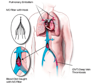
The Inferior vena cava is the largest vein of your body. It is the major channel connecting the veins of your leg and connecting them to the heart. The inferior vena cava carries all the impure de-oxygenated blood from your lower limbs as well as the kidneys back to your heart for purification. During DVT, clots form in the veins of your leg. In some patients these clots can migrate from the veins of your leg through the inferior vena cava and to your heart. From the heart these clots can migrate to the arteries of your lung (called the pulmonary artery) resulting in a condition called as pulmonary embolism (PE) or pulmonary thromboembolism (PTE). Due to large clots in the lung arteries, patients will devel difficulty in breathing or a drop in blood pressure. In rare cases, massive pulmonary embolism can also pose a risk to life.

The IVC filter is like an umbrella-shaped device which is placed in the inferior vena cava. It is a special device made up of metal which is placed either through the veins of your leg or through the neck veins into the inferior vena cava. Multiple shapes and types of inferior vena cava filters are available in the market. Consider the IVC filter to be a filtration mechanism wherein large clots from the deep veins of the leg are prevented from migrating to the heart and arteries of the lung. These clots get entrapped within the twines and prongs of the filter preventing upward migration. Therefore, an IVC filter is placed to prevent pulmonary thromboembolism.
An IVC filter does not have to be placed in all cases of DVT and pulmonary embolism.
The conventional and standard protocol treatment option for both DVT and pulmonary embolism is only medical management in the form of anticoagulation. Anticoagulants are drugs which are given to prevent further clot propagation and clot migration. In most cases only injectable anticoagulants followed by oral anticoagulants is the standard treatment for both DVT and pulmonary embolism. However, when the patient is not responding to anticoagulation or when there is massive DVT involving the complete lower limb venous system, newer treatment options like mechanical and pharmacomechanical thrombectomy to suck out the entire thrombus can be warranted. In cases of pulmonary thromboembolism there despite medical management the patient continues to have low saturation or low blood pressure, in those cases the clot from the pulmonary arteries can either be sucked out using special devices (called thrombectomy) or special drugs can be placed right within the thrombus through the special catheters called catheter directed thrombolysis (CDT).
Details of these procedures and whether you are a candidate for these procedures is dependent on case to case basis.
It has significantly reduced over the past few years due to the development of newer drugs and agents. However certain important indications of IVC filter are as follows:
Another new indication of IVC filter placement is prior to mechanical thrombectomy for massive iliofemoral deep venous thrombosis cases. In certain patients with massive DVT when the clot needs to be sucked out, an IVC filter is temporarily placed so that during the surgery and during manipulation no clot migrates into the lungs.
When can the IVC filter be removed?
Newly available IVC filters are all retrievable. The disadvantage of an IVC filter is that the patient needs to be on long-term or lifelong anticoagulation. Therefore, it is always advisable that the IVC filter be removed. However, depending on the indication of filter placement and on case to case basis, the timing of IVC filter removal needs to be decided in joint conjunction with your primary treating doctor.
How is the IVC filter removed?
Just like placement, the retrieval of the IVC filter can also be done under local anaesthesia. However, for difficult cases, sedation may be required. Most cases are done through the veins of your neck. However, in complex cases a combined groin as well as neck access may be used. Filters that are generally placed within one month can easily be removed without any difficulty. However, long-standing filters which are placed more than 1 to 3 months prior, pose a challenge. A prior CT scan may be required to localize the exact location of the prongs of the filter and the presence of any associated clot.
Depending on the complexity and the timing of filter placement, sometimes an IVC filter removal may be extremely difficult requiring multiple operators, refined techniques and special devices like forceps and balloons.