Septal Defects

ReivewsByCardiac

Medically Reviewed By Dr. Meghav Shah Updated on November 28, 2024 

Normally both the right and left atria i.e. the upper heart chambers are separated by an interatrial septum and the ventricles i.e. the lower heart chambers are separated by an interventricular septum. Any defect in these membranes is called septal defects.


Normally, the blood in the right side of the heart has deoxygenated (less oxygen) blood and the left side of the heart has oxygenated (more oxygen) blood. These septa normally prevent the mixing of the blood on the right side with the left side of the heart, thus maintaining the integrity of the normal blood circulation in the body.
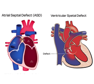
Septaldefectasdvsd

Types of Septal Defects

Subtypes of ASD

  • PFO or Patent Foramen Ovale - it is technically not a ASD. It is just that the septum primum and secundum fail to meet and close up. Normally it needs no treatment, but if patients have a stroke or recurrent migraine, then it may need to be closed.
  • Ostium Primum ASD - it occurs when the septum primum fails to fuse with the endocardial cushions and it is broadly associated with atrioventricular septal defects.
  • Ostium Secundum ASD - most common type of ASD wherein the septum secundum fails to occlude the ostium.
  • Sinus Venosus ASD - these occur near the place in the septum where the superior or the inferior vena cava drain in the right atria.
  • Coronary Sinus ASD - it occurs when there is unroofing of the coronary sinus, leading to blood flow from the left atria to right atria via the coronary sinus.


Subtypes of VSD

  • Membranous VSD - the defect is present in the upper part of the ventricular septum. It is the most common VSD and accounts for more than 75% of VSDs.
  • Muscular VSD - the defect is usually present in the mid to lower part of the ventricular septum. These account for around 20-25% of VSDs.
  • Outlet VSD - in the form of subaortic or subpulmonic VSDs. This defect is present in the septum just before the aortic or pulmonary arteries arise.
  • Inlet VSD - This defect is present in the septum located close to the tricuspid or the mitral valves.
  • Post Infarct VSD - this occurs secondary to extensive myocardial infarction or heart attack, that causes thinning and eventually rupture of the septum causing a secondary VSD due to loss of blood supply to that part of the septum. This is a serious and life threatening condition.

Risk Factors

These septal defects are congenital, i.e. people are born with them since birth.


Exact cause for same is not known, but it is due to varied genetic and environmental reasons.


Certain medicine intake or addictions like smoking or alcohol consumption in pregnancy can be a causative factor.


Many a times these defects may be a part of a more generalized syndrome disorder, for eg. Holt Oram Syndrome, Downs Syndrome etc.


Rarely, as mentioned previously VSD can occur secondary to massive heart attacks or by iatrogenic injury i.e. injury during some heart operations.

Symptoms of ASD & VSD

ASD

  • Many a times they are asymptomatic in children and are diagnosed incidentally when Echocardiogram is done for other reasons.
  • In adults, they may present with tiredness, shortness of breath, palpitations or swelling over the legs. In late cases patients can have cyanosis i.e. bluish tinge of lips and ears and thickening and rounding of finger nails called clubbing.


VSD

  • Symptoms of VSD depend on its location and size of defect.
  • Very small VSDs are asymptomatic and diagnosed when physician hears a murmur or Echocardiogram is done for other reasons.
  • Bigger defects can cause symptoms of shortness of breath or tiredness, palpitations and swelling over the feet. The bigger the defect, earlier the symptoms arise.

Diagnosis

  • Electrocardiogram
  • Echocardiography with colour doppler is the go to diagnostic procedure to diagnose these defects and plan what intervention may be needed for same
  • Occasionally, TEE (transesophageal echocardiography), i.e. a echo probe is passed in the gullet to have closer look at the heart, to identify these septal defects and more importantly to plan the further treatment order
  • Cardiac catheterization is occasionally needed to check the feasibility of the procedure to close the defects by mainly checking the pressures in the lungs and to check oxygen levels in the various heart chambers

Management & Treatment


Medicines 

Water tablets or diuretics are mainly for symptom relief if there is shortness of breath or swelling over the feet.


Occasionally beta blockers are given if there are symptoms of Palpitations or fast heart beating.


Also, in certain cases, medicines may be given to reduce the pressures in lungs.


Definitive Procedure

If amenable via percutaneous device closure i.e. putting a device through the groin arteries to close the defect is the preferred procedure. It can be done occasionally without general anesthetic as well. If successful, patients are discharged home the next day.


If device closure is not possible, then an open heart surgery can be done to close the defect.

Complications If Left Untreated

If untreated, these defects can cause irreversible damage to the heart and lungs called as Eisenmenger syndrome, and if this gets established these defects are deemed inoperable.


This occurs because there is increased blood flow to the right heart chambers because of the defect and subsequently more blood flows into the lungs. Over the time secondary to increased blood flow in lungs, the pressures in the lungs increases and this raised pressures becomes irreversible even when the defect is closed. On the contrary it may be of more harm than good if defects are closed post Eisenmengerisation.


These patients have a very poor quality of life, they turn blue secondary to more deoxygenated blood flowing in the body, are very lethargic and short of breath and have gross swelling over the feet.

Conclusion

ASD and VSD are usually congenital defects and need to be identified and treated early. Any child complaining of tiredness or shortness of breath should see a child physician and if deemed likely to have these conditions, they should be referred for an Echocardiogram. If diagnosed on same, these patients should be referred to pediatric cardiologists for consideration of closure in the form of Percutaneous device closure or open heart surgery.

Meet Our Consultants

Dr.AnkurPic
Dr. Ankur Phatarpekar

Structural & Interventional Cardiologist
MD, DM (Cardiology), FSCAI

Location - Dadar

View profile
Dr Meghav Images
Dr. Meghav Shah

Structural & Interventional Cardiologist
MD, DM, DNB (Cardiology)

Location - Ghatkopar

View profile