VascularSpecialistHospitalinMumbaiMobileBanner HVSHospitals

Our Services

We offer advanced care for a comprehensive range of services to diagnose, treat, and manage vascular conditions at HVS hospitals in mumbai.

Vascular Specialist Department

Vescular Overview New

Vascular Services at HVS Hospitals in mumbai are specialised services which are generally offered via small clinics across the country. Our Vascular department is an attempt to elevate the services with provision for best infrastructure including fully equipped ICU and dedicated specialists.

Vascular techniques in modern medicine are gaining recognition for being effective and less risky. At HVS hospitals, we are introducing the gold standards treatment in fully equipped hospital facility to ensure best results for patients.

  • Arterial Interventions
  • Vein care & Deep Venous Interventions
  • Neuro Interventions
  • Vascular Surgery
  • Hepatobillary & Portal Interventions
  • Onco Interventions
  • Dialysis Access and Nephrology IR
  • Vascular Diagnosis
  • Reproductive Health

Meet Our Consultants

Our dedicated team of vascular specialists are here to provide you with world-class care. With extensive experience and specialized expertise, our consultants are committed to delivering personalized and comprehensive treatment plans.

Doctor Bhavesh
Dr. Bhavesh Popat

Vascular & Interventional Radiologist
MD, FVIR, EBIR

Location - Dadar

View profile
Karan New Doctor
Dr. Karan Anandpara

Vascular & Interventional Radiologist
MD, FVIR, EDIR, EBIR

Location - Ghatkopar

View profile

Technology

Harnessing the latest advancements in medical technology, our Vascular Department offers state-of-the-art diagnostic and treatment options to ensure precise, effective, and minimally invasive care for all vascular conditions.

Case Study

Explore our success stories and detailed case studies to see how our expert team and advanced treatments have transformed the lives of patients with complex vascular conditions.
Case Study

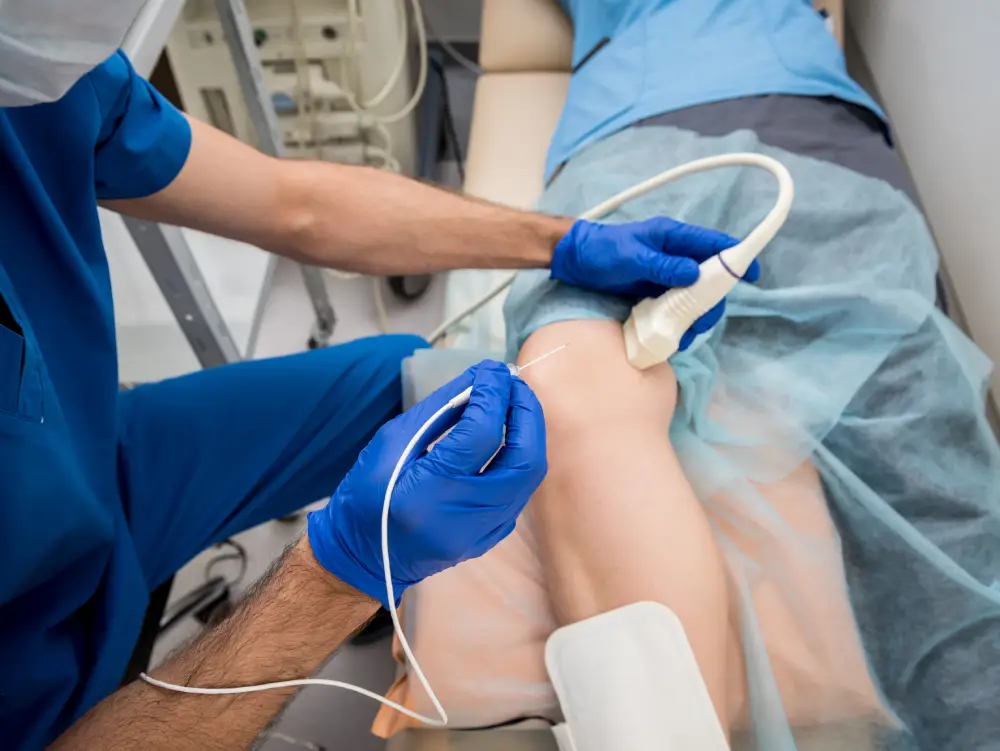
Geniculate Artery Embolization

Procedure/Surgery Name: Geniculate Artery embolization for Recurrent Hemarthrosis

Doctors: Dr Bhavesh Arun Popat, Dr Karan M Anandpara

Pre-Op Details & Diagnosis

A 72 years old male

Comorbidities: Morbidly obese, poorly controlled diabetes, hypertensive, previous H/O CABG.

Complaints: Bilateral knee pain, joint aches and soreness (left>right knee).

Pain after joint overuse.

Reduced physical activity and quality of life.

Local knee tenderness +.

X ray bilateral knee s/o bilateral osteoarthritic changes (left more than right).

H/O left sided recurrent hemarthrosis for which percutaneous USG guided aspiration of fluid done outside.

USG local part suggestive of left sided hemarthrosis – hyperechoeic fluid with septations in the left knee joint cavity.

MRI done s/o changes of severe osteoarthritis with left sided hemarthrosis

Pain not relieved by conservative medical management i.e. by anti-inflammatory drugs or corticosteroids.

Not relieved by intra-articular steroid injection.

Not a candidate for joint replacement/arthroplasty in view of significant medical comorbidities, contraindications for general anaesthesia and patient’s refusal for knee arthroplasty.

Treatment Plan

Planned for Left sided Geniculate Artery Embolization in view of recurrent hemarthrosis and left joint pain.

Details about the Procedure/Surgery:

Procedure under local anesthesia. Groin prepared.

Left Common Femoral Artery antegrade access.

DSA Angiogram done which showed hypertrophied superior and inferior medial geniculate arteries.

Synovial vascular blush noted.

Super selective cannulation of the superior and inferior geniculate arteries done using Progreat microcatheter.

Embolization done with PVA particles.

Post Procedure Details:

Post procedure DSA showed reduction in the synovial vascularity. Reduced arterial blush noted.

Sheath removed by manual compression.

Patient discharged on same day.

Significant reduction of pain on 1 and 3 month follow up.

Conclusion

Geniculate artery embolization (GAE) reduces pain of osteoarthritis by decreasing the vascular supply to the hypertrophied and inflamed synovium and has a role in cases of recurrent hemarthrosis.

It is safe, effective and minimally invasive.

No cuts, no stitches are required.

It can be Performed under local anesthesia.

No general anaesthesia needed.

It is a day care procedure with a same day discharge.

Read More
Case Study 2

Endovascular And Percutaneous

Procedure/Surgery Name: Endovascular Glue Embolization of lower limb/leg Arterio-Venous Malformation (AVM)

Doctors: Dr. Bhavesh Arun Popat, Dr. Karan M Anandpara

Pre-Op Details & Diagnosis

A 29 year old female

  • Progressively increasing painful swelling on anterior aspect of right distal leg of 4 months duration
  • No skin changes
  • Minimal right distal leg edema
  • Tender on palpation +
  • Distal pulses (ATA, DPA and PTA) palpable +

Investigation

  • Local Ultrasound and Doppler – echogenic ill defined soft tissue area with few serpiginous vascular channels within s/o vascular malformation
  • MRI – confirmed the findings of USG; arterial feeders with intralesional nidus noted s/o likely arterio-venous malformation (AVM)

Angiography Findings:

  • Abnormal leash of vessels
  • Internal nidus
  • Atleast 4 arterial feeders from anterior tibial artery
  • Early draining vein – washout from accompanying anterior tibial veins
  • Distal main anterior tibial artery and DPA shows slow, but antegrade adequate distal flow

Diagnosis

Complex arterio-veneous malformation (avm) of lower limb/leg with multiple arterial feeders from anterior tibial artery.

Treatment Plan

  • Staged; combined ultrasound and fluoroscopic percutaneous and sos endovascular glue embolisation
  • Followed by surgical excision

Procedure Details

  • Percutaneous USG guided access taken
  • Catheter in the right popliteal artery via left CFA access
  • Endovascular contrast injection to localize the nidus and to monitor residual lesion
  • Following this, multiple percutaneous access taken to target various components of the AVM
  • Manual hand compression given to prevent inadvertent reflux into the draining vein
  • Endovascular embolization attempted, but ultimately was not done due to high probability of reflux into the main anterior tibial artery due to intra-nidus vascular communication. Therefore, percutaneous embolization done with Glue

Post Procedure DSA

  • Near complete exclusion of the AVM
  • Glue cast visible

Conclusion

  • Essential to classify vascular malformations appropriately – Venous, lymphatic, AVM, combined vascular malformations
  • Set ‘realistic expectations’
  • Multiple procedures
  • Difficult to treat completely
  • Team Work
Read More
Case Study 4

Endovascular Aneurysm Repair

A 55 years male patient with history of sudden abdominal pain followed by syncope was rushed to the hospital. CT abdomen + angiogram s/o fusiform dilatation of the abdominal aorta. Eccentric thrombus along the left lateral wall, showing further extension along the aortic bifurcation to involve bilateral common iliac arteries may represent thrombosed aneurysm/ intramural hematoma.

Investigation

The findings most likely represent a ruptured & thrombosed fusiform aortic aneurysm. On receiving patient in ICU he was in hypovolemic shock with poor general condition. Blood pressure on admission was 80/40 mmHg, with a heart rate of 120/min. Physical examination of the abdomen was soft with tenderness in the left lower quadrant. No pulsatile mass was present. Femoral pulses were palpable symmetrically. Nor Adrenaline infusion started. Routine investigations sent and volume resuscitation was done with Blood transfusion followed by Fresh Frozen Plasma. Hb was 6.5 with an INR of 1.2. Rest routine reports were within acceptable range.

Procedure Details

Patient was stabilized and shifted to Cath Lab for Abdominal Aortic Stenting.

She underwent Abdominal Stenting with Graft by a team of Interventional Radiologists & Cardiologists. Post procedure PCV transfused with Hb of 8.0. There was no further drop in Hb and rest of hospital stay was uneventful and patient discharged on POD 5.

Read More
Case Study 3

EVAR 70 yrs

A 70 years male was admitted and diagnosed with abdominal aorta aneurysm for planned abdominal aortic aneurysm stenting. He had history of low back pain for 2 years. His Follow up scan showed increase in size of Aortic Aneurysm with rapid progression of disease in Right Iliac artery. Considering his advanced age and co morbidities, patient was not willing for surgery and opted for minimal intervention.

After detailed evaluation patient underwent Abdominal Aortic stent grafting under general anaesthesia. Patient tolerated the procedure well and shifted to the ICU. All invasive lines removed gradually. He was treated with IV Antibiotics, LMWH, DAPT, Statin, Beta blocker and other supportive measures. The patient's postoperative course was uneventful and he was discharged on postoperative Day 4 with no renal impairment.

Read More

Testimonials

Google logoStar rating
My mother underwent an Aortic valve repair procedure at the hospital. Given her age and her condition, we were not sure whether to go ahead with the procedure. However, Dr. Phatarphekar was extremely kind, pleasant, helpful and approachable. He and his team explained to us every minute detail related to the procedure and cleared all our doubts and inhibitions.

- Mangesh Thorat

HandWrittenTestimonials

Locations

location description

Heart & Vascular Superspeciality Hospitals - Ghatkopar

3rd Floor, Silver Point, LBS Marg, Kasturi Park, Maneklal Estate, Ghatkopar West, Mumbai - 400086

HVS Symbiosis Superspeciality Hospitals - Dadar

4th, Chabildas Rd, Dadar West, Dadar, Mumbai - 400028

Frequently Asked Questions